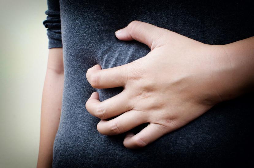
Hernia is a protrusion of the bowels or adjacent organs/tissue through a defect or weakness in the abdominal wall. Typically, a hernia occurs with a part of the intestine protruding or hanging out through an opening formed in the abdominal wall. Smoking, diabetes, age and obesity also contribute toward this condition.
Incisional hernia is a type of hernia that develops at the site of an incision from a previous surgery. It may develop shortly after the surgery or years later. They are especially susceptible three to six months following the procedure, when the tissues are healing from the incision. Strenuous activity, substantial weight gain, or pregnancy can cause excessive stress on the healing abdominal tissue and should be avoided during this healing window. Prevalence reported ranges from 0.5% to 13.9% for most abdominal surgeries.
Symptoms and signs
A patient with incisional hernia could feel deep discomfort and pain. Especially while coughing, lifting heavy objects or standing for long hours. This pain results from the pressure of the organ or tissue that is pushing its way through the defect in the abdominal wall. As the condition worsens, the pain increases.
Apart from feeling pain, the patient may also be able to see the hernia. One may observe a lump in the abdomen or groin. This lump may enlarge over time.
The following symptoms and signs of an incisional hernia include:
- Fever
- Infection
- Bulging (palpable lump and/or mass on surgical scar)
- Visible protrusion (see internal segment poking out of surgical wound)
- Pain
- Pressure
- Aching
- Swelling
- Foul-smelling drainage
- Redness and/or red streaks (sign of infection)
- Bowel obstruction (strangulation of intestines)
Signs of this type of hernia are found during the examination process, such as bulging near the surgical scar, visible protrusion of internal tissue, organ, intestine, muscle, and/or fatty contents, and bleeding. The most common hernias exhibit symptoms and signs that resemble each other.
How is incisional hernia diagnosed?
A physical examination is performed to diagnose an incisional hernia. Upon diagnoses, the patient will have to consult with a surgeon for further treatment.
In some cases, it may be appropriate radiological exams to diagnose incisional hernia as:
- Abdominal ultrasound can be used to detect fascial defects as well as differentiate between an incarcerated incisional hernia and a solid mass;
- Abdominal and pelvic CT scan is excellent in the detection of incisional hernias and characterization of involved viscera. CT scan is particularly useful in diagnosing acute incarceration in morbidly obese patients in whom physical examination is difficult and unreliable.
Treatments
Treatments for an incisional hernia require surgery. The hernia will not heal on its own without surgery. Surgery is necessary to push the protruding tissue back in place, remove any scar tissue, and adhere a surgical mesh on the hernia’s opening to prevent recurrence.
Hernia must be treated immediately. If it is not repaired at the earliest, more risks and complications may result in the future. An untreated hernia only gets worse increasing the levels of discomfort and pain. Patients must understand the severity of an untreated incisional hernia to avoid more serious complications such as strangulation of the intestine, which can lead to tissue death of this important internal membrane. As result of which last clinical complication, patient will need an urgent surgery.
The following two types of surgeries are recommended treatment options to repair an incisional hernia:
- Open hernia repair: most common surgical technique involves making a skin incision over the affected site to make a hernia repair.
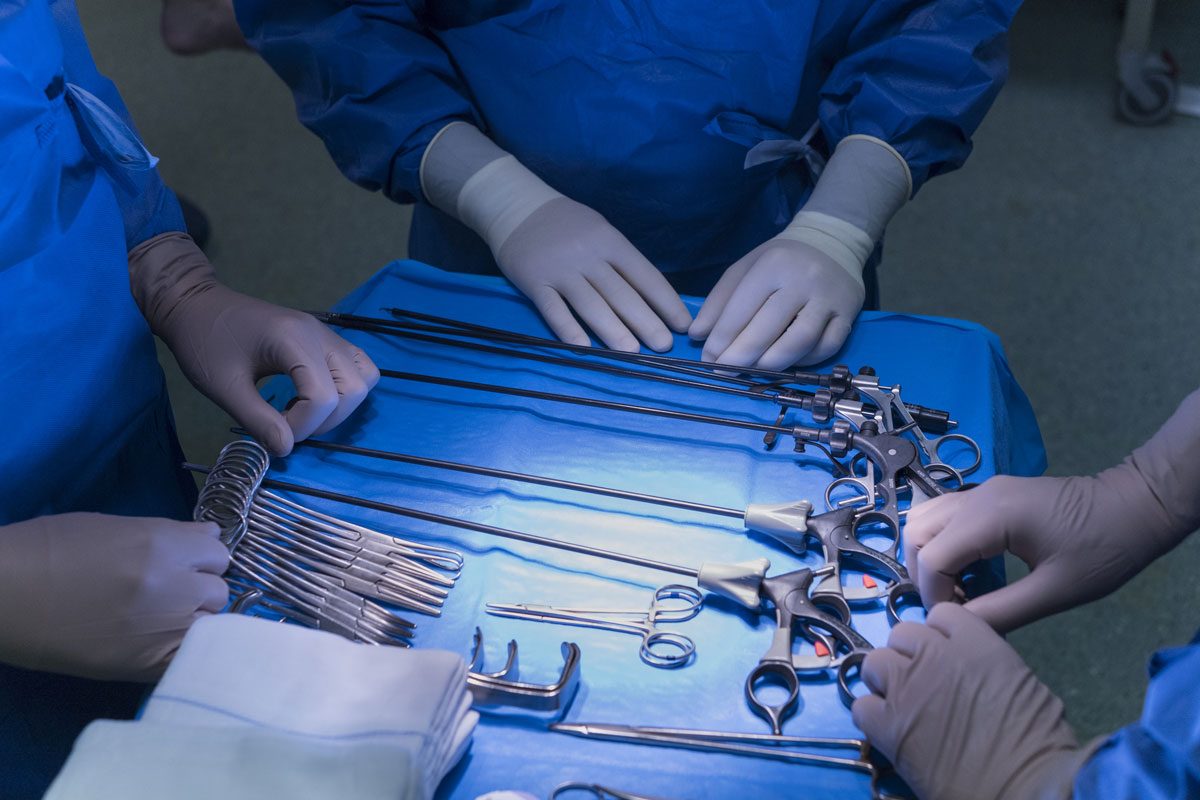
- Laparoscopic hernia repair: A less invasive surgery that uses a small tube with a camera to repair the hernia. This surgical device is inserted into small incisions of the abdomen, where the surgeon can watch this procedure on a monitor.
Postsurgical Complications
Complications after surgical hernia repair may occur in up to 50% of cases, depending on surgical technique and the status of the hernia sac vasculature. Approximately one-half of these complications may require surgical reintervention, and accurate diagnosis at CT scan is necessary for optimal patient treatment.
- Hernia Recurrence: the most common complication after hernia repair, usually occurring 2–3 years after surgery. The prevalence varies with the type of repair:
- up to 30% of cases after open surgery without mesh placement;
- up to 10% after open surgery with mesh placement
- up to 7.5% after laparoscopic surgery
- a small percentage occur 5 or more years after surgery, usually related to the aging of tissues and the weakening of muscles.
- Fluid Collections: occur frequently (≈ 17% of cases) in the immediate postoperative period after hernia repair. These collections usually contain serous fluid (seromas) or blood products (hematomas). Most seromas resolve without manipulation within 30 days; aspiration may be indicated if the collection persists for more than 6 weeks, steadily grows, produces symptoms, or is suspected to be infected. Imaging-guided aspiration or drainage may be problematic for large collections located under the mesh.
- Infection: infected postoperative fluid collections occur in 1%–5% of patients. These complications tend to occur more frequently in older female patients, especially after surgical repair of strangulated and incarcerated hernias. Moreover, they tend to manifest early in the postoperative period (<2 weeks after surgery) and constitute an important risk factor for hernia recurrence. Infected fluid collections may involve subcutaneous (superficial) or mesh-surrounding (deep) tissues. Differentiation is important because superficial infections are managed conservatively, whereas deep infections require intervention such as percutaneous drainage or prosthesis removal.
- Mesh-related complications:
- Inflammatory reactions may lead to fibrosis of tissues surrounding the mesh;
- Mesh shrinkage may also occur;
- Intraperitoneal adhesions may develop, predisposing to small bowel obstruction;
- Migration of mesh within the abdominal wall (less frequently).


